Cell
- A cell is the smallest unit that is capable of performing life functions.
- Structural & functional unit of Life.
- Basic unit of Biological activity.
Prokaryotic Cell:
- Do not have structures surrounded by membranes
- Few internal structures
- Examples: One-celled organisms, Bacteria
Eukaryotic Cell:
- Contain organelles surrounded by membranes
- Most living organisms
Difference between Prokaryote and Eukaryote cell
| Prokaryotic Cell | Eukaryotic cell | |
| Nucleus | Absent. DNA found as nucleoid. | Present. Surrounded by nuclear membrane. |
| Nuclelolus | Absent | Present |
| Subcellular organelles | Absent | Present |
| Transport | Absent | Present |
| Ribosome | 70 S | 80 S |
| Cell wall | Present | Absent |
| Cell division | Binary fission | Mitosis, Meiosis |
| Size | Smaller (1-10 microm) | Larger (10-100 microm) |
| Energy | Mitochondria absent; enzymes bound to membrane. | Mitochondrial enzymes |
| Examples | bacteria, mycoplasma | Animal cell, plant cell, fungi |
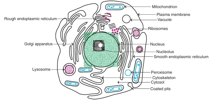 Fig: Structure of a human cell
Subcellular Organelles
Cell contains various organized structures such as Nucleus, Mitochondria, Lysosomes, Golgi complex, Endoplasmic reticulum, peroxisomes etc.
Nucleus
- It is present in all cells of body except mature erythrocytes
- It directs cell activities
- Separated from cytoplasm by nuclear membrane
- It contains genetic material – DNA
- Functions: DNA Replication and RNA Synthesis (transcription) takes place inside the nucleus
Nuclear Membrane:
- Surrounds nucleus
- Made of two layers
- Openings allow material to enter and leave nucleus
Chromosomes:
- Inside nucleus, DNA combines with histone proteins to form chromatin
- Chromatin threads assemble & condense to form chromosomes
- Number of chromosomes is specific to each species. DNA of each human cell contains 23 pairs of chromosomes, a total of 46.
- Chromosomes contain instructions for traits & characteristics
Nucleolus:
- In some cells, portion of nucleus may be seen as light shaded area called nucleolus
- Rich in enzymes: DNA polymerases, RNA polymerases etc
- RNA processing and ribosome synthesis takes place at nucleolus.
Cytoplasm
- Gel-like mixture
- Region of cell outside the nucleus
- Functions: Protein synthesis, Glycolysis, Glycogen metabolism, HMP Shunt, Fatty acid synthesis, Cholesterol synthesis, heme synthesis (part), urea synthesis (part), pyrimidine synthesis (part), purine synthesis
Endoplasmic Reticulum (ER)
- Network of Membrane enclosed spaces or cisternae
- Under electron microscope, reticular arrangement have railway track appearance.
- When cells are fractioned, ER is disrupted to form microsomes.
- Functions: 1. Smooth type ER: lacks ribosome : Lipid synthesis and calcium storage
- 2. Rough type ER: Ribosome embedded in surface : Protein Synthesis. Protein part of glycoproteins and lipoproteins are synthesized in ER.
- 3. Microsomal cytochrome p450: Detoxification of many drugs
- 4. Ethanol oxidation, synthesis of cholesterol (partial)
Ribosomes
- Each cell contains thousands of ribosomes
- They are Factories of Protein synthesis.
- Found on ER & floating throughout the cell
Mitochondria
- Spherical, oval or rod like bodies. Erythrocytes lack mitochondria.
- It Produces energy through chemical reactions – breaking down fats & carbohydrates: Power house of the cell
- Mitochondrial DNA is transmitted by cytoplasmic inheritance (maternal)
- Functions: 1. Cristae contains Electron transport chain – ATP generation takes place
- 2. Mitochondrial cytochrome p450 system involved in steroidogenesis.
- 3. TCA cycle, gluconeogenesis (part), beta oxidation of fatty acids, ketone bodies production
- 4. Heme synthesis (part), urea synthesis (part), pyrimidine synthesis (part),
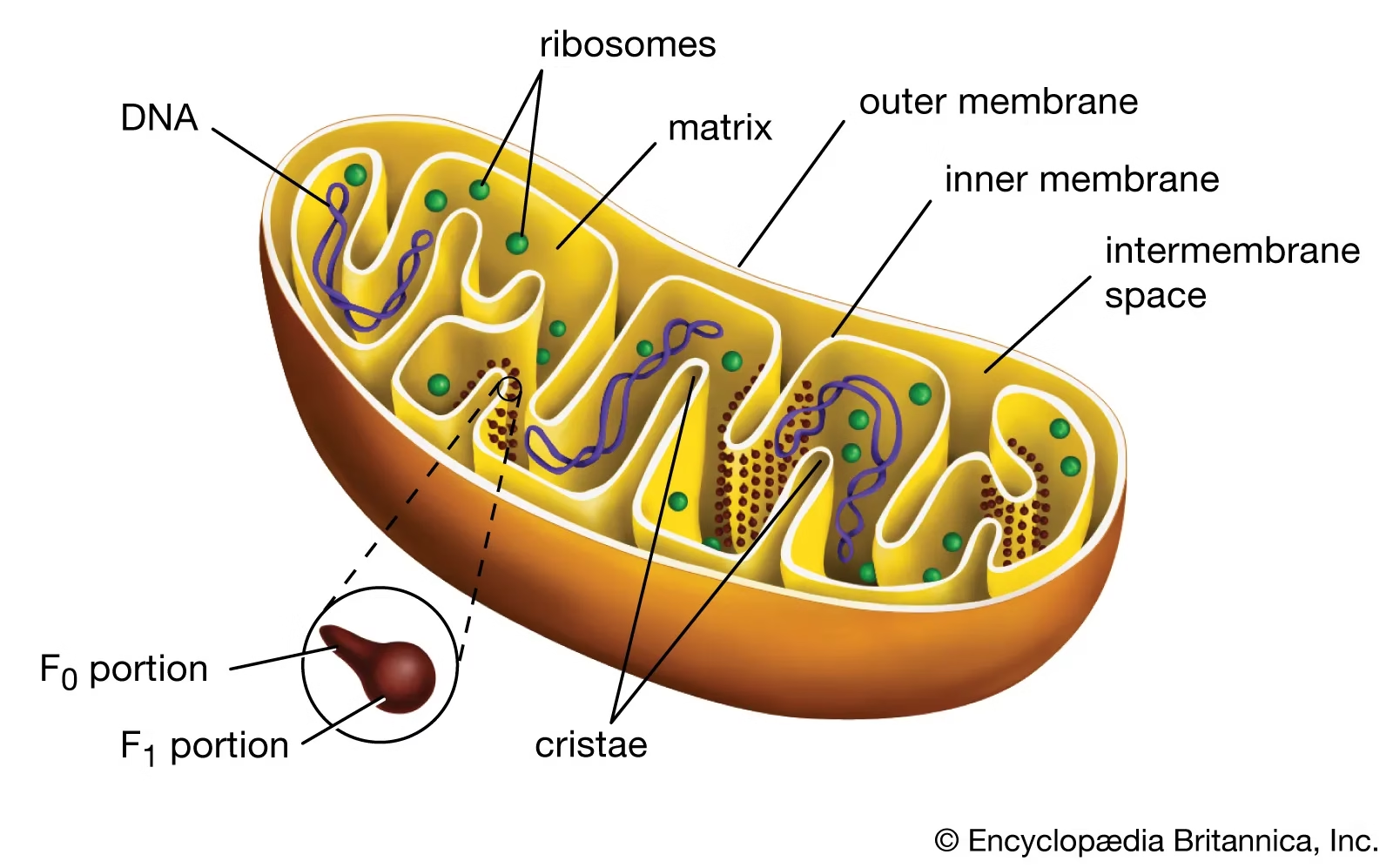
Fig: Mitochondria
Components of Mitochondria:
- Outer membrane
- Inner membrane
- cristae
- Intermembrane space
- Matrix
Golgi Bodies or Golgi Complex
- Network of Membrane vesicles- It acts as converging area of ER.
- Protein ‘packaging plant’: Newly synthesized proteins are first sorted
- Glycoproteins are transported from ER to golgi apparatus for temporary storage.
Lysosomes
- Bags of enzymes called as Digestive ‘plant’ for proteins, fats, and carbohydrates:
- Digestive tract of the cell: Transports undigested material to cell membrane for removal
- Contain many degradative enzymes such as 1. Lipid hydrolyzing enzymes: Lipases
- 2. Polysaccharide hydrolyzing enzymes: Alpha glucosidase, beta galactosidase, hyaluronidase
- 3. Protein hydrolyzing enzymes: Cathepsins, Collagenases, elastases
- 4. Nucleic acid hydrolyzing enzymes: Ribonucleases
- Functions: Degradation of proteins, carbohydrates, lipids, nucleotides
- Maintains cellular components in dynamic state
- Inclusion Cell (I-cell) disease : Rare condition where lysosomes lack enzymes
Peroxisomes
- Microbodies: Major role in fee radical scavenging
- Catalase and peroxidase enzyme: protects cell from toxic effects of H2O2 and destroy unwanted peroxides
- Oxidation of long chain Fatty Acids occurs in peroxisomes
Marker Enzymes
Some enzymes are present in certain organelles called as marker enzymes.
| Subcellular Organelle | Marker Enzyme |
| Mitochondria | ATP Synthase |
| Lysosomes | Cathepsin, Acid phosphatase |
| Golgi complex | Galactosyl transferase |
| Microsomes | Glucose-6-phosphatase |
| Cytoplasm | Lactate Dehydrogenase |
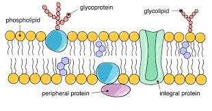
Cell membrane/ Plasma Membrane
- Plasma membrane separates the cell from external environment.
- It has Highly selective permeability properties- Entry and exit of compounds are regulated.
- Membranes are made up of lipids, proteins and small amount of carbohydrates.
- Membranes are made of a double layer of lipid molecules containing proteins which are embedded in them.
Lipids:
- The major membrane lipids are phospholipids.
- These are amphipathic molecules containing both hydrophilic or polar (water soluble) groups oriented towards extracellular side and hydrophobic or non-polar (water insoluble) groups towards cytoplasmic side.
- Phospholipids in cell membrane are organized to form bilayers. The phospholipid bilayer is the basic structure of membranes.
- The cell membrane also contains equal amount of cholesterol.
Proteins:
- Proteins in membranes occur in two different ways.
- Some proteins are located in the interior of the membrane and are called integral membrane proteins. (attached by hydrophobic bonds or van der Waals forces)
- Others are located in the outer surface of the membranes and are known as peripheral membrane proteins. (tethered to membrane by covalent linkages with membrane lipids).
- Some integral membrane proteins span the the whole lipid bilayer called as Transmembrane proteins.
- Transmembrane proteins can serve as:
- a) Channels b) Carriers c) Pumps d) Receptors for hormones, growth factors, neurotransmitters
Carbohydrates:
The extracellular surface of the plasma membrane contains small amounts of carbohydrate structures known as glycocalyx. Carbohydrates are present as glycoproteins and glycolipids. Carbohydrates play an important role in cell signaling and in identifying and interacting with other cells.
Fluid mosaic structure:
Membranes are organized into a structure known as the fluid mosaic model. It is called so because membrane proteins float in a sea of lipids. This model of the cell membrane was proposed by Singer and Nicholson in 1972.
- Thickness 5-8 nm.
- Cell membrane is Asymmetric : due to irregular proteins
- Appearance of ceramic or mosaic tile
- But unlike fixed tile: membrane freely changes; hence called as Fluid mosaic.
Factors affecting Membrane fluidity:
- Maintained by length of hydrocarbon chain, degree of unsaturation and nature of polar head group of lipids.
- Trans fatty acids (TFA) decrease the fluidity of membranes.
- Unsaturated cis fatty acids increase the fluidity
- Cholesterol content also alters fluidity o the membrane.
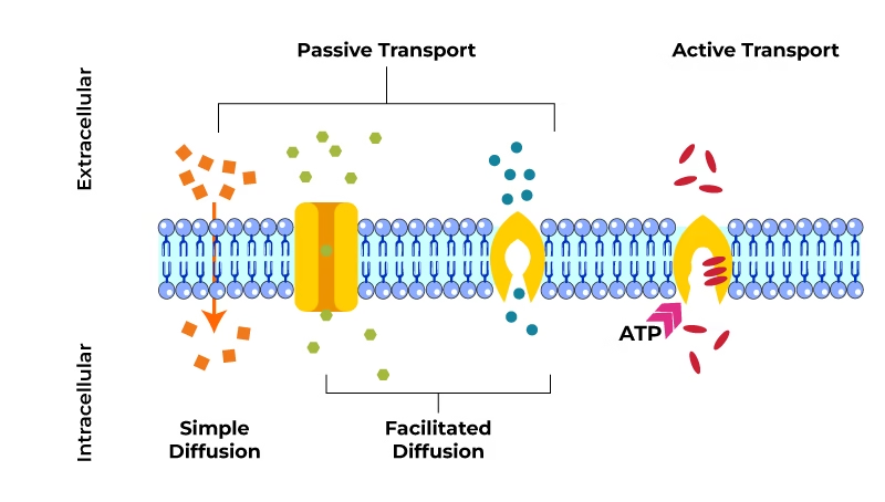
Transport Across Biological Membranes
Membrane restricts unwanted molecules and permits entry of molecules necessary for cellular metabolism.
Transport mechanisms are classified as:
- Passive Transport
- Active Transport
Passive Transport
Here, substances move across the cell membrane without any energy expenditure by the cell. It includes diffusion and osmosis.
Diffusion:
Simple/ Passive Diffusion:
- Along the concentration gradient.
- Occurs from higher to lower concentration.
- Passage of water & gases
- Does not require energy.
![]()
Facilitated Diffusion:
- Along the concentration gradient
- No energy needed. Can operate bidirectionally.
- Mediation of Carrier or Transport proteins: Transport proteins facilitate diffusion across membranes
- Carrier molecules exist in Ping and pong states. In Pong state- active sites are exposed to exterior, where solutes bind. In Ping state- active sites is changed to interior, which cause release of solute molecules.
Osmosis:
The diffusion of water from an area of high concentration of water molecules (high water potential, low osmotic pressure) to an area of low concentration of water (low water potential, high osmotic pressure) across a partially permeable membrane.
Active transport
When substances are transported against their chemical and electrical gradient requiring energy, the process is called active transport.
- Against a concentration gradient
- Requires energy
- Carrier mediated process
- Requires specialized integral proteins called transporters
The main types are
- Primary active transport
- Secondary active transport
- Carrier type transport
- Vesicular transport
Primary Active Transport
Directly uses energy obtained from hydrolysis of ATP
Examples: i) Sodium–potassium (Na+–K+) pump or Na+– K+ ATPase.
- It drives sodium ions from intracellular to extracellular fluid and potassium ions in the opposite direction.
- Responsible for maintenance of High K+ and low Na+ concentration in the cells.
ii) Calcium (Ca2+) pump: ATP dependent Calcium pump functions to regulate muscle contraction.
Secondary Active Transport
Uses electrochemical gradient across plasma membrane as its energy source, instead of ATP hydrolysis
Examples: i) Na+ dependent secondary active transport: Glucose and amino acids are reabsorbed from PCT or reabsorbed from intestinal lumen if Na binds to protein
ii) Na+– Ca+ Exchanger
iii) Iodide pump in thyroid gland
Carrier Type Transport
It involves proteins that act as transporters.
Carriers are transport proteins that bind to ions/molecules and moves from one side of membrane to the other.
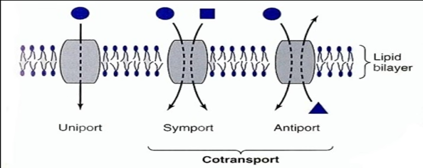 Fig: Carrier type transport: Classified by direction of movement and whether one or more unique molecules are moved
- Uniport -system moves one type of molecule bidirectionally Ex: Glucose transporter in most of the cells
- Cotransport– systems transfer one solute dependent upon simultaneous or sequential transfer of another solute. It is of two types
- Symport – moves solutes in same direction (Ex: Na+-sugar transporters use in ORT or Na+-amino acid transporters)
- Antiport – moves two molecules in opposite directions (Ex: Na+ in and Ca2+ out)’
-
Vesicular Transport or Transcytosis
Large molecules are transported across the cell membrane by endocytosis and exocytosis.
- Endocytosis:
- Cell internalize extracellular macromolecules, to form endocytic vesicle.
- Phagocytosis— “Cell eating” Ex: Engulfment of large extracellular particles like bacteria, dead tissue, foreign particles by macrophages.
- Pinocytosis– “Cell drinking”
- Receptor-mediated endocytosis- Selective pinocytosis Ex: Uptake of LDL, Clathrin coated pit absorption of cholesterol
- Exocytosis: or Reverse Pinocytosis
- Secretory vesicles move towards and fuse with cell membrane, where contents vesicles are externalized.
- Example: Cellular secretion
Mnemonics
1. Components of Cell Membrane (Fluid Mosaic Model)
Mnemonic: “Phat People Can Make Fun Friends”
P – Phospholipids
P – Proteins (Integral & Peripheral)
C – Cholesterol
M – Membrane carbohydrates (Glycolipids, Glycoproteins)
F – Fluid mosaic
F – Flexibility
2. Functions of Cell Membrane
Mnemonic: “PETERS”
P – Protection
E – Endocytosis & Exocytosis
T – Transport (Selective permeability)
E – Enzymatic activity
R – Receptor for signaling
S – Structural support
3. Types of Membrane Transport
Mnemonic: “P-FATE” (Passive and Facilitated transport are FATE-ful)
P – Passive transport
F – Facilitated diffusion
A – Active transport
T – Transcytosis (Endo- & Exocytosis)
E – Endocytosis (Pinocytosis, Phagocytosis)
4. Types of Membrane Proteins
Mnemonic: “TRICE”
T – Transport proteins
R – Receptor proteins
I – Integral proteins
C – Cell recognition proteins
E – Enzymatic proteins
5. Differences: Active vs Passive Transport
Mnemonic for Active: “NEEDS ATP”
-
N – Needs Energy (ATP)
-
A – Against gradient
-
T – Transport proteins needed
-
P – Pumps (Na+/K+ pump)
Mnemonic for Passive: “HIGH to LOW, No ATP Flow”
6. Types of Endocytosis
Mnemonic: “PPR” – Please Pick Right
P – Phagocytosis (“cell eating”)
P – Pinocytosis (“cell drinking”)
R – Receptor-mediated endocytosis