Cell
- A cell is the smallest unit that is capable of performing life functions.
- Structural & functional unit of Life.
- Basic unit of Biological activity.
Prokaryotic Cell:
- Do not have structures surrounded by membranes
- Few internal structures
- Examples: One-celled organisms, Bacteria
Eukaryotic Cell:
- Contain organelles surrounded by membranes
- Most living organisms
Difference between prokaryote and eukaryote cell:
| Prokaryotic Cell | Eukaryotic cell | |
| Nucleus | Absent. DNA found as nucleoid. | Present. Surrounded by nuclear membrane. |
| Nuclelolus | Absent | Present |
| Subcellular organelles | Absent | Present |
| Transport | Absent | Present |
| Ribosome | 70 S | 80 S |
| Cell wall | Present | Absent |
| Cell division | Binary fission | Mitosis, Meiosis |
| Size | Smaller (1-10 microm) | Larger (10-100 microm) |
| Energy | Mitochondria absent; enzymes bound to membrane. | Mitochondrial enzymes |
| Examples | bacteria, mycoplasma | Animal cell, plant cell, fungi
|
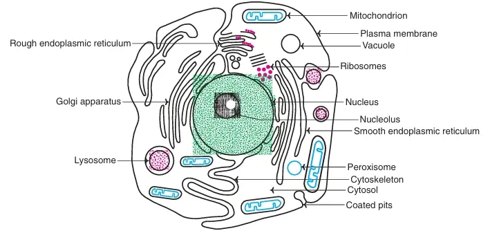 Fig: Structure of a human cell
Cellular Organelles:
Nucleus:
- Directs cell activities
- Separated from cytoplasm by nuclear membrane
- Contains genetic material – DNA
Nuclear Membrane:
- Surrounds nucleus
- Made of two layers
- Openings allow material to enter and leave nucleus
Chromosomes:
- In nucleus
- Made of DNA.
- DNA associated with basic protein (histones): Nucleosomes
- Assembly of nucleosomes: Chromatin fibres of chromosomes
- Contain instructions for traits & characteristics
Nucleolus:
- Inside nucleus
- Contains RNA to build proteins
Nucleolus:
- Ground material of nucleus
- Rich in enzymes: DNA polymerases, RNA polymerases etc
Cytoplasm:
- Gel-like mixture
- Surrounded by cell membrane
Endoplasmic Reticulum:
- Membrane enclosed spaces
- Smooth type: lacks ribosome : Lipid synthesis
- Rough type (pictured): ribosome embedded in surface : Protein Synthesis
- Rough ER is disrupted to form Microsomes.
Ribosomes:
- Each cell contains thousands
- Factories of Protein synthesis.
- Found on ER & floating throughout the cell
Mitochondria:
- Produces energy through chemical reactions – breaking down fats & carbohydrates
- Power house of the cell
- Recycles and decomposes proteins, fats, and carbohydrates
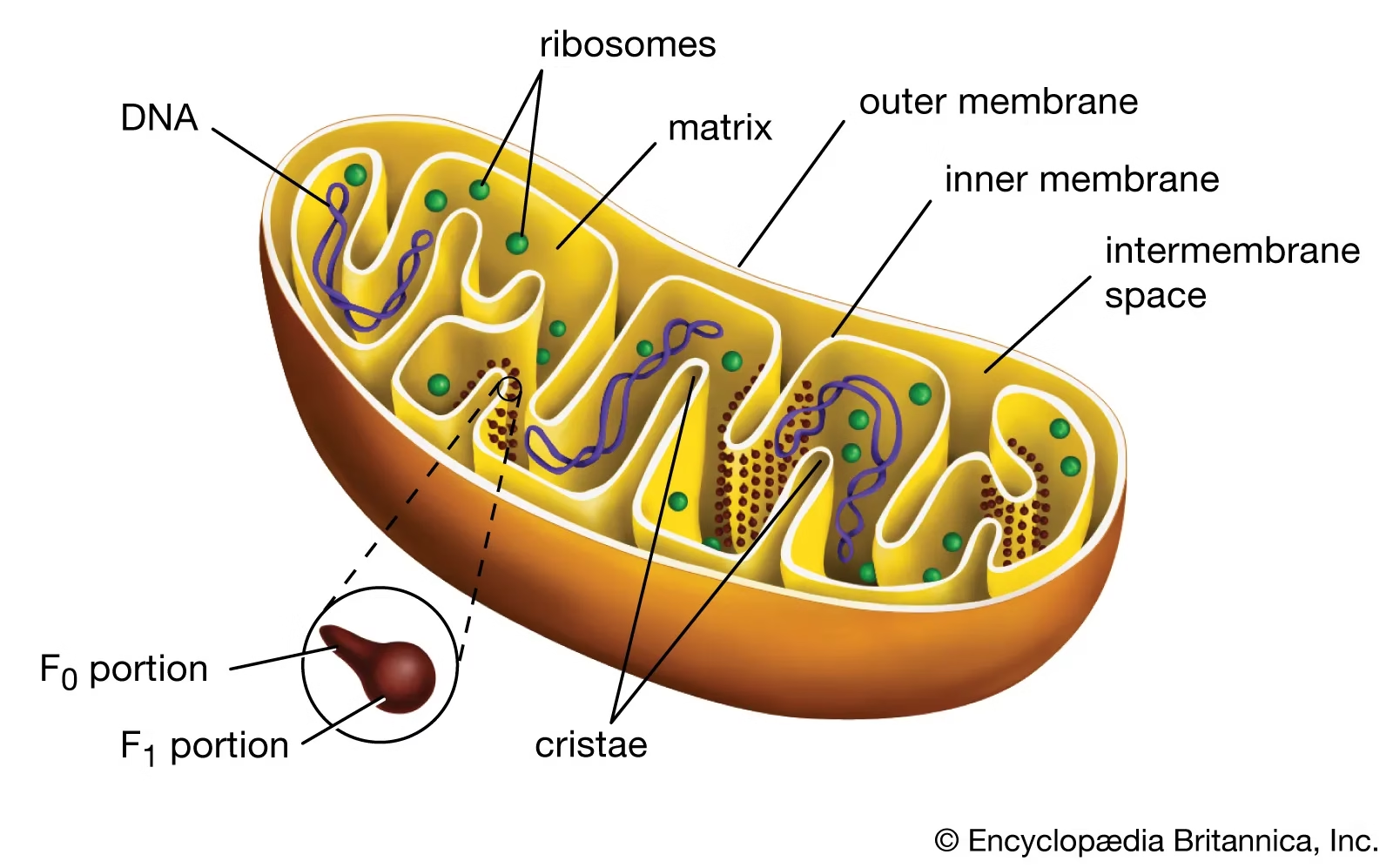
Fig: Mitochondria
Components of mitochondria:
- Outer membrane
- Inner membrane
- cristae
- Intermembrane space
- Matrix
Golgi Bodies:
- Membrane vesicles: Dictyosomes
- Protein ‘packaging plant’
- Move materials within the cell
- Move materials out of the cell
Lysosomes:
- Digestive ‘plant’ for proteins, fats, and carbohydrates
- Transports undigested material to cell membrane for removal
- Cell breaks down if lysosome explodes
- Contain many degradative enzymes such as acid hydrolases g.
- Glucosidase, cathepsins, lipases, ribonucleases
- Digestive tract of the cell
- Maintains cellular components in dynamic state
Peroxisomes:
- Microbodies
- Catalase enzyme: protects cell from toxic effects of H2O2.
- Oxidation of long chain FA
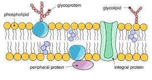
Cell membrane
Membranes are made of a double layer of lipid molecules containing proteins which are embedded in them.
Lipids:
- The major membrane lipids are phospholipids. These are amphipathic molecules containing both hydrophilic or polar (water soluble) and hydrophobic or non-polar (water insoluble) groups.
- The cell membrane also contains equal amounts of cholesterol.
Proteins
- Proteins in membranes occur in two different ways. Some proteins are located in the interior of the membrane and are called integral membrane proteins.
- Others are located in the outer surface of the membranes and are known as peripheral membrane proteins.
- Transmembrane proteins serve as:
- a) Channels b) Carriers c) Pumps d) Receptors
Carbohydrates:
The extracellular surface of the plasma membrane contains small amounts of carbohydrate structures known as glycocalyx.
Fluid mosaic structure:
Membranes are organised into a structure known as the fluid mosaic model. It is called so because membrane proteins float in a sea of lipids. This model of the cell membrane was proposed by Singer and Nicholson in 1972.
- Thickness 5-8 nm.
- Lipid bilayer
- Asymmetric : irregular proteins
- Appearance of ceramic or mosaic tile
- But unlike fixed tile: membrane freely changes; hence Fluid mosaic.
Phospholipid Bilayer:
Phospholipids can form bilayers.
2 layers of phospholipids with hydrophobic tails protected inside by the hydrophilic heads.
The phospholipid bilayer is the basic structure of membranes.
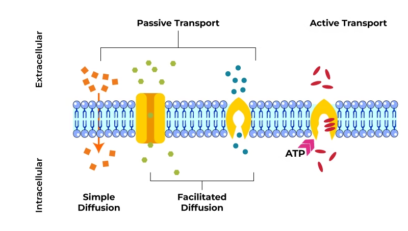
Transport Across Biological Membranes
Passive Transport
Here, substances move across the cell membrane without any energy expenditure by the cell. It includes diffusion and osmosis.
Diffusion:
Simple/ Passive Diffusion:
- Depends on concentration gradient
- Passage of water & gases
- Does not require energy.
![]()
Facilitated Diffusion:
- Along the concentration gradient
- No energy needed.
- Mediation of carrier or Transport proteins: transport proteins facilitate diffusion across membranes
Osmosis:
The diffusion of water from an area of high concentration of water molecules (high water potential, low osmotic pressure) to an area of low concentration of water (low water potential, high osmotic pressure) across a partially permeable membrane.
Active transport:
When substances are transported against their chemical and electrical gradient requiring energy, the process is called active transport.
- Against a concentration gradient
- Requires energy
- Carrier mediated process
The main types are
- Primary active transport
- Secondary active transport
- Carrier transport
- Vesicular transport
1. Primary Active Transport
i) Sodium–potassium (Na+–K+) pump:
Responsible for maintenance of High K+ and low Na+ concentration in the cells.
Integral plasma membrane protein, Enzyme Na+– K+ ATP ase.
High cellular K+ is required for optimal glycolysis and for protein synthesis.
ii) Calcium (Ca2+) pump
iii) Potassium–hydrogen (K+–H+) pump
2. Secondary Active Transport
3. Carrier Type Transport
-
- Uniporters
- Symporters
- Antiporters
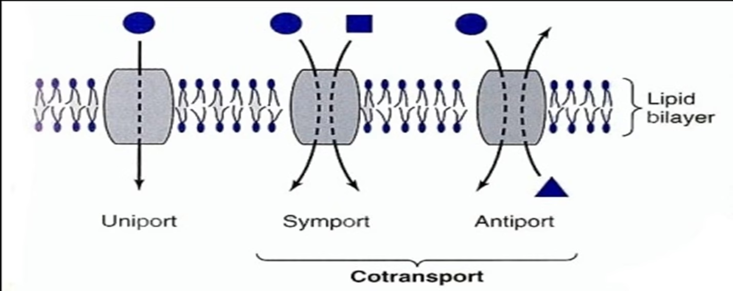 Fig: Carrier type transport: Classified by direction of movement and whether one or more unique molecules are moved
- Uniport -system moves one type of molecule bidirectionally
- Cotransport- systems transfer one solute dependent upon simultaneous or sequential transfer of another solute
- Symport – moves solutes in same direction (g., Na+-sugar transporters use in ORT or Na+-amino acid transporters)
- Antiport – moves two molecules in opposite directions (g., Na+ in and Ca2+ out)
- Vesicular Transport or Transcytosis
Many substances are transported across the cell membrane by endocytosis and exocytosis.
- Endocytosis—
- Phagocytosis— “Cell eating”
- Pinocytosis– “Cell drinking”
- Receptor-mediated endocytosis-specific particles, recognition.
- Exocytosis—Cellular secretion
- i) Endocytosis: pinocytosis, phagocytosis
- ii) Exocytosis